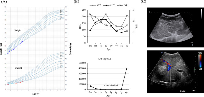
FIGURE 2.

Clinical parameters and images of the patient. (A) Growth chart, (B) laboratory findings including transaminase, INR, and AFP, (C) liver ultrasound images showing cirrhotic liver with nodules at the last visit (upper panel), a large hyperechoic mass newly appeared at diagnosis of HCC (lower panel)
