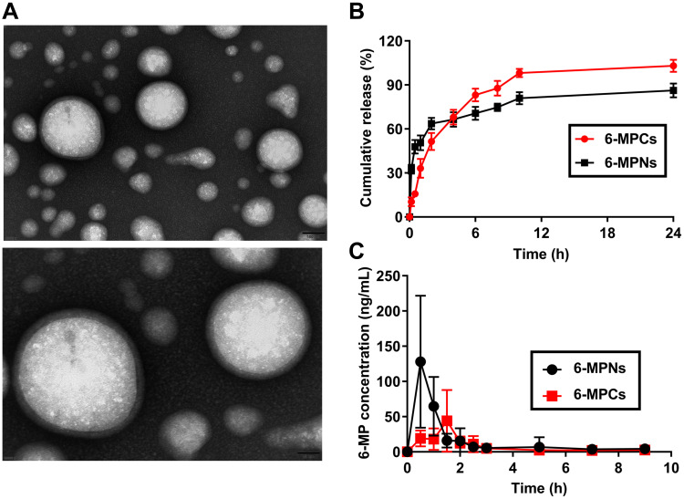
Figure 1.
Characterization of 6-MPNs in vitro and in vivo. The morphology of 6-MPNs measured by Transmission Electron Microscope (TEM) (A). (B) In vitro release of 6-MP from 6-MPNs and 6-MPCs in PBS containing 0.02% Tween 20. (C) In vivo plasma concentrations (ng/mL) of 6-MP vs Time (h) profiles after a single oral administration of 6-MPNs or 6-MPCs in SD rats.

