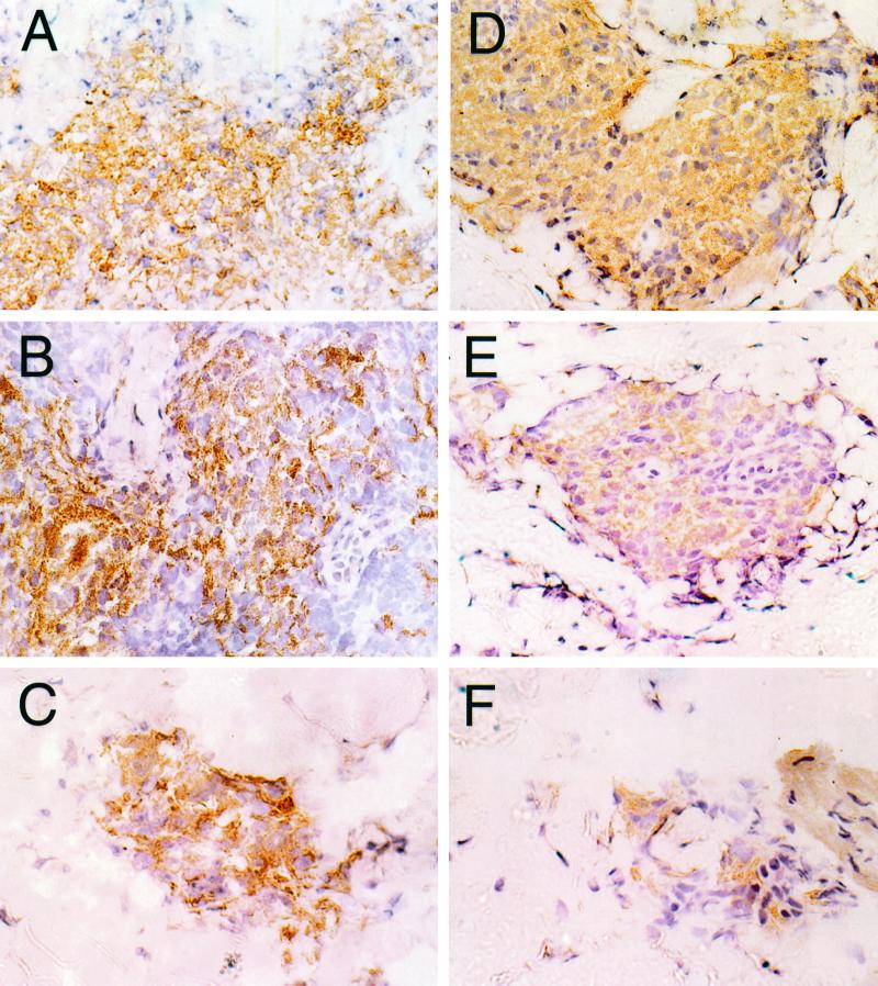
FIG. 1.
Photomicrographs of tissue sections stained with immunoperoxidase for IFN-γ and IL-12 expression in a BT patient (patient 380; BT-RR, 40× objective). (A) IFN-γ staining in a reactional granuloma in a biopsy taken at day 0. (B) A high level of IFN-γ expression is seen and appears unchanged at day 7. (C) Fewer cells are present in a smaller granuloma at day 28, and the level of IFN-γ expression has decreased compared with expression in the day 0 and 7 biopsies. IL-12 is strongly expressed in granulomas in biopsies taken at day 0 (D), and a similar level of positive staining is seen at day 7 (E). (F) The granuloma size and level of positive staining have decreased by day 28.

