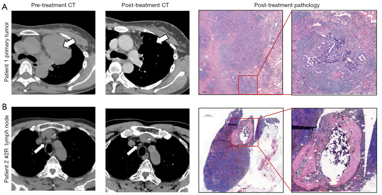
Figure 2.
Typical cases of radiological and pathological response to neoadjuvant immunochemotherapy. (A, left 2 figures) Thorax CT image of the primary tumor from a 56-year-old female non-smoker patient diagnosed with stage IIIB (T4N2M0) lung adenocarcinoma before and after two cycles of neoadjuvant pembrolizumab + pemetrexed disodium + carboplatin. The white arrow represented the position of the primary tumor before and after neoadjuvant treatment. The patient then underwent left upper lobectomy. (A, right 2 figures) Representative sections of the primary tumor surgical resected specimen (HE staining, 10× and 40×) with 30% residual tumor cells in the regression bed. (B, left 2 figures) Thorax CT image of the #2R lymph node from a 63-year-old male patient with stage IIIC (T4N3M0) lung squamous cell carcinoma who received 10 cycles of neoadjuvant sintilimab + cisplatin + abraxane. The write arrow illustrates the swollen #2R lymph node before the treatment and its shrinkage after the treatment. The patient then underwent right upper lobectomy. (B, right 2 figures) Representative sections of the surgical resected #2R lymph node specimens (HE staining, 10× and 40×), showing a regression bed inside the lymph node, which is characterized by the features of cell death (cholesterol clefts and interstitial foamy macrophages are obvious in the regression bed). CT, computed tomography.

