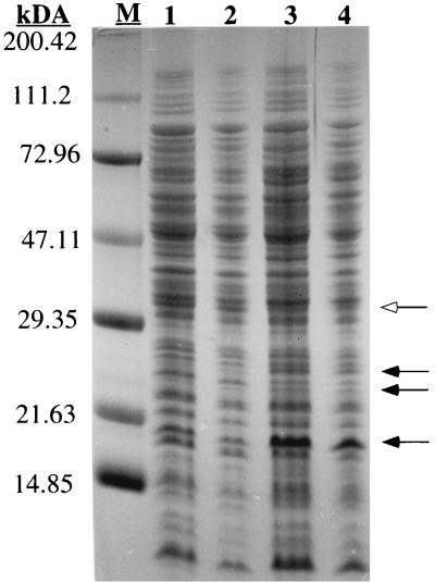
FIG. 4.
Surface-associated proteins. Surface proteins from S. aureus ATCC 35556 strains were released with 1% SDS, separated on an SDS–15% polyacrylamide gel, and stained with Coomassie blue. Lanes: 1 and 2, extracts from the wild type; 3 and 4, extracts from dltA mutant. Protein extracts (50 or 25 μl) were applied to lanes 1 and 3 or 2 and 4, respectively. The masses of protein markers (M) are indicated on the left. Protein bands with different intensities in the two strains are indicated by closed (more pronounced in the wild type) and open (more pronounced in the dltA mutant) arrows.

