FIG. 3.

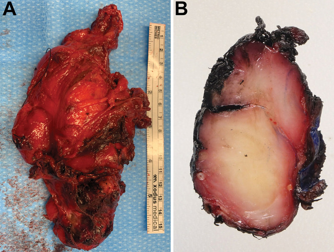
A: Photograph of the excised mass showing irregular, red-brown, ragged, firm tumor tissue. B: A cut section of the excised tissue demonstrating the firm homogeneous tissue architecture and areas of focal hemorrhage.

A: Photograph of the excised mass showing irregular, red-brown, ragged, firm tumor tissue. B: A cut section of the excised tissue demonstrating the firm homogeneous tissue architecture and areas of focal hemorrhage.