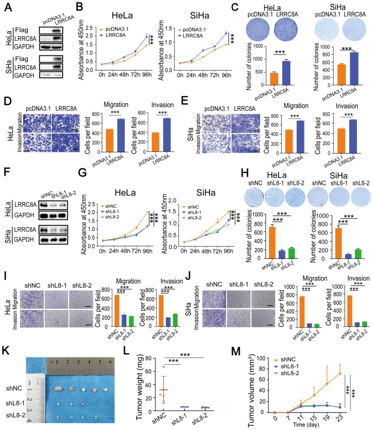
Figure 2.
LRRC8A promotes tumorigenesis of CC in vitro and in vivo. (A) Western blot analysis of LRRC8A protein expression in CC cells with LRRC8A overexpression. (B) Cell growth curves of CC cells with or without LRRC8A overexpression by CCK-8 assays. (C) LRRC8A contributes to the proliferation of HeLa and SiHa cells as measured by colony formation assays. (D, E) Cell migration and invasion capacities of CC cells with LRRC8A overexpression. Scale bar, 200 μm. (F) LRRC8A knockdown was confirmed in HeLa and SiHa cells by western blot. (G) Effects of LRRC8A knockdown on abilities of CC cells' proliferation. (H) Colony formation assays were performed in CC cells upon LRRC8A knockdown. (I, J) Transwell assays detecting migration and invasion of CC cells with LRRC8A depletion. Scale bar, 200 μm. (K-M) Effects of LRRC8A knockdown on tumor weight and volume in the subcutaneous xenograft nude mouse model. Data are presented as means ± S.D: ***P < 0.001.

