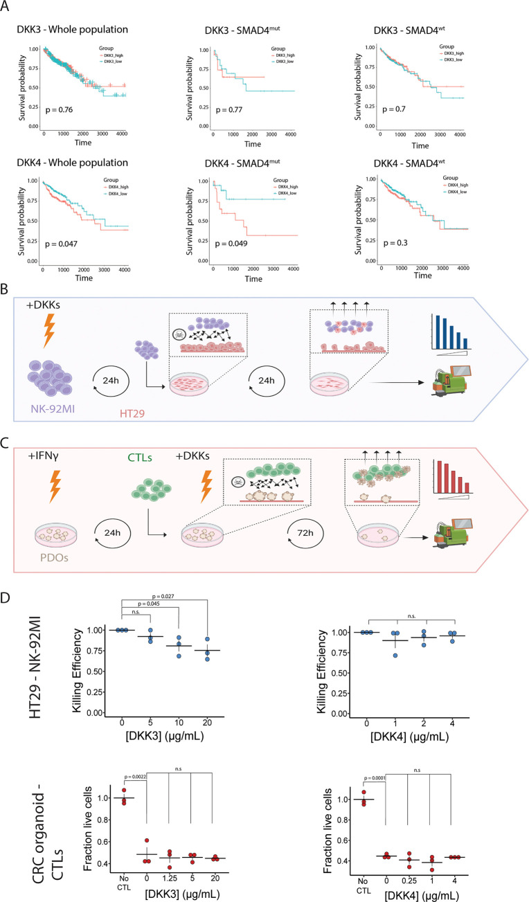
Figure 3.
DKK3 reduces the killing capacity of NK cells for CRC cells in vitro. (A) Kaplan–Meier plots showing the effect of DKK3 (top) and DKK4 (bottom) expression on survival probability in the TCGA-COAD cohort. Left panels include the whole cohort, middle panels show patients with mutations in SMAD4, and right panels show patients without SMAD4 mutations. (B) Schematic overview of killing assays using CRC cells and NK cells. NK-92MI cells are incubated for 24h with recombinant DKK-proteins after which they are co-cultured with HT29 cells. After 24h of coculture, non-adherent cells are removed and adherent cells are dissociated and counted by flow cytometry (C) Schematic overview of killing assays using patient-derived CRC tumor organoids and matched CTLs. Patient derived organoids (PDOs) are incubated with IFNγ for 24h after which they are cocultured with CTLs in combination with recombinant DKK proteins. After 72h, non-adherent cells are removed and adherent cells are dissociated and measured by flow cytometry (D) Dot plots showing the effect of DKK3 and DKK4 on the antitumor capacity of NK cells (top) and CTLs (bottom). No CTL indicates the control condition, where no CTLs are added to the tumor organoids; n = 3 independent replicates; error bars represent SEM. n.s: not significant.

