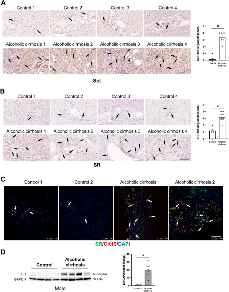
Fig. 2.
Sct and SR immunoreactivity increased in liver sections of patients with alcoholic cirrhosis compared to healthy control livers. A Sct and B SR immunoreactivity in human liver sections was examined by immunohistochemistry (4 healthy and 4 patients with alcoholic cirrhosis, from n = 8 independent samples per group. Representative images are shown at orig. magn., 20X, scale bar = 100 μm. Immunohistochemical quantification of Sct and SR in human liver sections. Data are mean ± SEM of 5 random fields. *p < 0.05 vs. respective healthy control livers. Each dot represents one value in data set. Black arrows indicate bile ducts positive for Sct or SR, whereas white arrows indicate bile ducts positive for SR. C Immunofluorescence in liver sections co-stained with CK19 (SR staining from 2 healthy controls and 2 patients with alcoholic cirrhosis). Representative images are shown at orig. magn. 20X, scale bar: 100 μm. D By immunoblots, SR protein levels increased in total liver from patients with alcoholic cirrhosis (n = 4) compared to human healthy controls (n = 4). *p < 0.05 vs. healthy control livers. Data are mean ± SEM of three separate immunoblots from total liver from patients with alcoholic cirrhosis compared to human healthy controls

