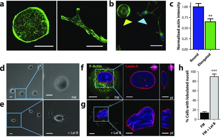
Figure 2.
Nuclear shape requires intact F-actin structures. (a to e) Fluorescence confocal and phase contrast micrographs of T47D cells after 48 h exposure to FM and FM + 250 µM of actin-depolymerizing drug Latruculin B, respectively, for 48 h. Cells are stained for F-actin (green), Lamin A (red), and nuclear DNA (blue). Scale bar, 10 µm. Panels to the right of f and g show zoomed in xy and yz cross-sections of the left panels. Scale bar 5 µm. (h) Percentage of cells featuring a lobulated nucleus 48 h after exposure to FM (black) or FM + Lat B (gray). Numbers of examined cells are n = 92 andn = 49 for FM and FM + Lat B stimulated cells, respectively.

