
Figure 2.
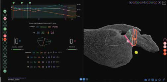
Web-based 3D imaging exploration (VIDAA platform). Left: Morphological parameters (e.g., diameters) of 2D contours along the centerline characterizing the left atrial appendage (LAA) anatomy and range of most appropriate devices to implant. Right: 3D visualization of the LAA anatomy in a 3D wire-frame mesh format, together with the LAA centerline (white), several 2D contours and some anatomical landmarks (pink, orange, and yellow small spheres corresponding to the ostium, the landing zone, and the circumflex artery, respectively) relevant for the device implantation.