
Figure 4. Confocal microscopy of fluorescently labeled hyalocytes.
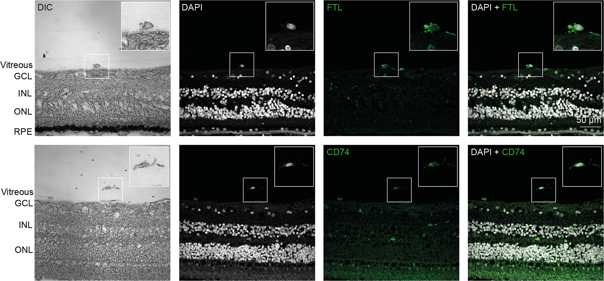
TOP: A preretinal hyalocyte close to the inner limiting membrane is shown. The hyalocyte was identified by its location via differential interference contrast (DIC) and a nuclear counterstain with DAPI to investigate the expression of the ferritin light chain (FTL). BOTTOM: A free hyalocyte with filopodia is shown. The hyalocyte was identified by its location via differential interference contrast and a nuclear counterstain with DAPI to investigate the expression of CD74. GCL = ganglion cell layer, INL = inner nuclear layer, ONL = outer nuclear layer, RPE = retinal pigment epithelium. Reproduced from [30], licensed under CC-BY 4.0 (http://creativecommons.org/licenses/by/4.0/)