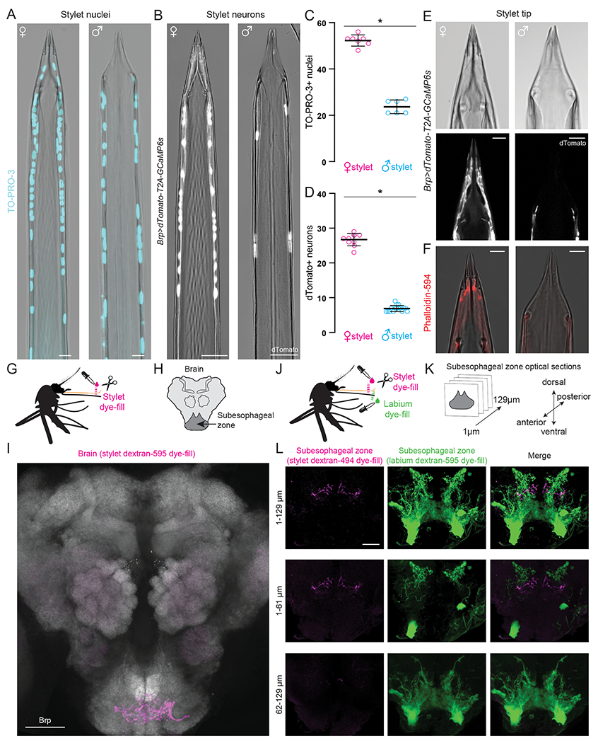
Figure 3. Sensory Neurons in the Female Stylet are Sexually Dimorphic and Project to a Unique Subesophageal Zone Region.

(A,B) Confocal image with transmitted light overlay of TO-PRO-3 nuclear staining (cyan) in wild-type female (A, left) and male (A, right) stylets, and dTomato expression (gray) in Brp>dTomato-T2A-GCaMP6s female (B, left) and male (B, right) stylets.
(C,D) Average # of TO-PRO-3 nuclei/stylet for most distal 300 μm (C, N=7 females, N=6 males), and dTomato neurons/stylet (D, N=10 females, N=16 males). Each dot denotes 1 animal (mean ± SD, * p < 0.05 Mann-Whitney test).
(E) Confocal image of transmitted light (top) and dTomato (gray, bottom) in Brp>dTomato-T2A-GCaMP6s female (left) and male (right) stylet tip.
(F) Confocal image with transmitted light overlay of phalloidin-594 (red) staining in wild-type female (left) and male (right) stylets.
(G, J) Schematic of stylet (G) and double (J) dye-fill experiment set-up performed in (I) and (L), respectively.
(H, K) Schematic of mosquito brain region captured in (I), and subesophageal zone optical sections captured in (L).
(I) Stylet neuron projection pattern (magenta) revealed by dextran-595 dye-fill. Neuropil stained with anti-Drosophila Brp (gray).
(L) Optical subesophageal zone sections from most anterior (top row) to most posterior (bottom row) of stylet (left, magenta) and labium (middle, green) projection pattern revealed by dual dextran-494 and dextran-595 dye-fill.
Scale bar: 50 μm (I), 25 μm (A,B,L), 10 μm (E,F).
See Figure S2 for additional analysis of stylet sexual dimorphism and Video 2 for confocal Z-stacks of dual dye-fills.
