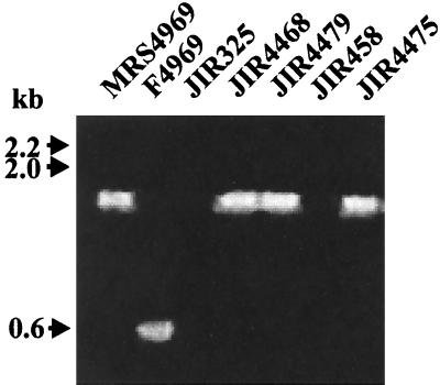
FIG. 1.
PCR analysis of transconjugants. Fresh cells from agar plates were resuspended in 100 μl of H2O and lysed in a microwave oven for 3 min. Three microliters of the supernatant was used in a standard PCR (50-μl volume) in a DNA thermal cycler (Perkin-Elmer) with 30 cycles of 30 s at 94°C, 30 s at 50°C, and 90 s at 72°C. The primers were cpeR (5′-CATCACCTAAGGACTGTTCT-3′) and cpeF (5′-TGTAGAATATGGATTTGGAAT-3′). The resultant products were separated by agarose gel electrophoresis. As shown, wild-type cpe-positive cells yielded a cpe band with a size of 544 bp, whereas a 1.7-kb band was present in strains with the cpeΩcatP region. Molecular sizes of the DNA markers are shown on the left.

