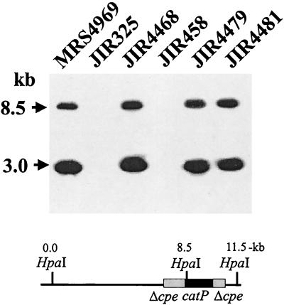
FIG. 2.
Southern blot of parent strains and transconjugants. C. perfringens DNA was isolated as described previously (14), digested with HpaI, separated by electrophoresis on a 1% agarose gel, and transferred by Southern blotting to positively charged nylon membranes (Roche Molecular Biochemicals). The membrane was then hybridized at 68°C overnight with a digoxigenin-labeled cpe probe. To prepare the probe, an ≈1.6-kb fragment containing the cpe gene and the regions ≈300-bp upstream and ≈200-bp downstream, was gel purified from EcoRI- and XbaI-digested pJRC200 DNA (14) and labeled by using a random-primed DNA labeling system (Boehringer Mannheim). After hybridization, the membrane was washed twice for 15 min each in wash solution (2× SSC [1× SSC is 0.15 M NaCl plus 0.015 M sodium citrate]–0.1% sodium dodecyl sulfate) at room temperature. The final washes were twice for 15 min each in 0.5× wash solution (0.5× SSC–0.1% sodium dodecyl sulfate) at 68°C. Hybridized probe was then detected by using a digoxigenin-chemiluminescence detection system with CSPD substrate (Roche Molecular Biochemicals). Molecular sizes of the DNA markers are shown on the left. The map shows the relevant cpeΩcatP region of pMRS4969.

