. 2022 Dec 15;11:e77945. doi: 10.7554/eLife.77945

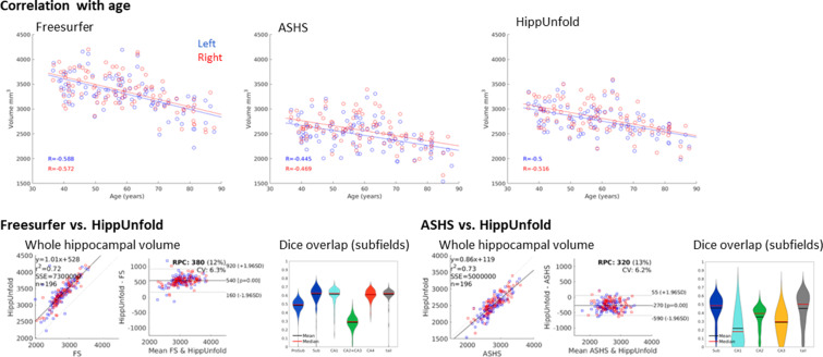
Figure 3. Out of sample performance of HippUnfold, Automatic Segmentation of Hippocampal Subfields (ASHS), and Freesurfer (FS7).

(A) Side-by-side comparison of results obtained from each method from one representative individual from the Human Connectome Project-Aging (HCP-A) datasets, which was not seen during training. (B) Quantitative comparison of subfield volumes (left) and age-related volume changes (right) between methods. For a full set of snapshots illustrating the differences between these methods, see Supplementary file 2, Supplementary file 3.

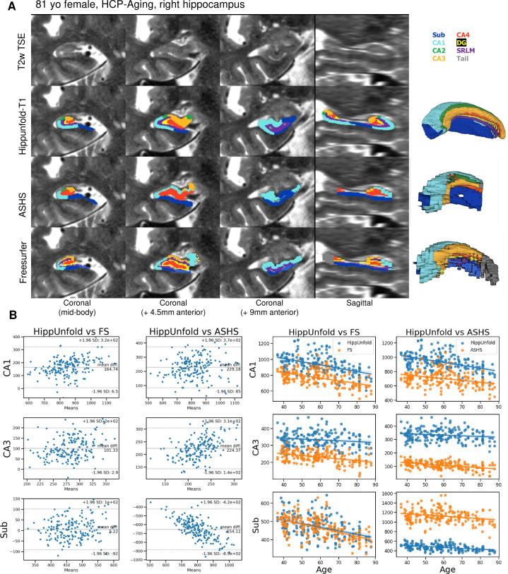
Figure 3.

Figure 3—figure supplement 1. Additional comparisons of results obtained from Freesurfer (FS7), Automatic Segmentation of Hippocampal Subfields (ASHS), and HippUnfold in 100 Human Connectome Project- Aging (HCP-A) subjects.

Figure 3—figure supplement 1.

All three methods showed a moderate correlation with age, as expected based on previous literature. Volumetric comparison of each method to HippUnfold directly revealed that there is a strong correlation between total hippocampal volumes obtained using HippUnfold and those obtained using FS7 or ASHS. HippUnfold and FS7 showed a moderate difference in overall volume (FS7 being on average 540 mm3 larger), whereas ASHS volumes were consistently smaller (by an average of 270 mm3). At the subfield level, using an unfolded subfield atlas from the corresponding method, there was relatively low Dice overlap between labels obtained using these three different methods in native space, which is likely driven by the gross volume differences between methods (i.e. which tissues are included or excluded prior to unfolding) since subfield definitions are nearly identical after unfolding.