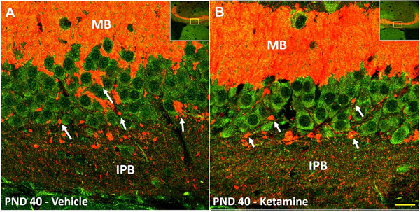
Fig. 4. Remaining IPB fibers likely make functional synapses with pyramidal layer neurons at PND40.
Immunofluorescence co-labeling of PSD-95 (green) and synaptoporin (red) in PND40 animal following neonatal vehicle (left panel) or ketamine (right panel), visualized with 60× objective. Multiple collaterals were observed emanating from the remaining IPB fibers as they migrate towards the MB. As these collaterals crossed the pyramidal layer, multiple large nodules (arrows) were detected in close proximity to PSD-95 positive pyramidal neurons, possibly en passant synapses, and also numerous small puncta around cell soma and basal dendrites. These histomorphological features of putative connections are indicative of functional synaptic connections between the presynaptic IPB terminals and CA3 pyramidal neurons. Insert is the 10× magnification image showing the location from which the 60× images were obtained. Scale bar – 20 μm. (For interpretation of the references to colour in this figure legend, the reader is referred to the web version of this article.)
IPB, infrapyramidal bundle; MB, main bundle; PND, postnatal day.

