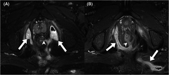
FIGURE 1.

Pelvic MRI of a 20‐year‐old man with CD for preoperative evaluation of the perianal fistula. (A) An axial T2‐weighted image with fat saturation presents a suprasphincteric fistula tract at the 3 o'clock position (arrowhead), with branching and abscess formation at bilateral ischioanal fossae (arrows). (B) a postcontrast axial T1‐weighted image of another fistula tract (arrowhead), with branching fistulae extending to the right ischioanal fossa and left gluteal region (arrows)
