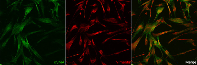
Fig. 2. Activated human induced pluripotent stem cell-derived cardiac fibroblasts.
Laser scanning confocal image of human induced pluripotent stem cell-derived cardiac fibroblasts activated by transforming growth factor-β stained with antibodies against α-smooth muscle actin (green) and vimentin (red). Every third to fourth cell in the left ventricle is a fibroblast. Most cardiac research has focused on the cardiomyocyte with research on cardiac fibroblasts proving more challenging. (Image provided by Ms Caitlin Hall, Institute of Cardiovascular Sciences, University of Birmingham).

