Figure 4.
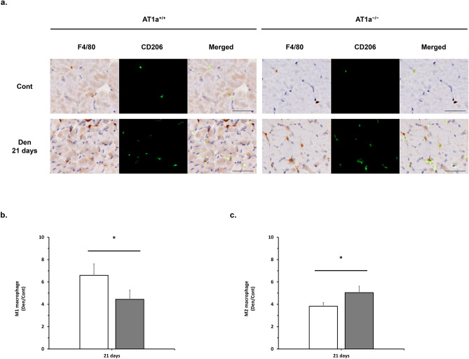
AT1a receptor loss modulated M1/M2 macrophage polarization in the denervated gastrocnemius muscle. (a) Immunostaining of gastrocnemius muscle in each AT1a+/+ and AT1a−/− mice at 21 days postdenervation. Immunostaining shows codistribution of M1 and M2 macrophages. Anti-F4/80 antibody binds M1 macrophages and M2 macrophages (brown color). Anti-CD206 antibody binds M2 macrophages (green fluorophore). CD206-/F4/80+ cells are M1 macrophages. CD206+/F4/80+ cells are M2 macrophages. Scale bar: 50 μm. (b, c) The degrees of infiltrated M1 macrophage (b) and M2 macrophage (c) were shown as the fold increase or decrease in the Den group compared with the Cont group in immunohistochemistry analysis. AT1a+/+-Cont group, n = 6; AT1a+/+-Den group, n = 7; AT1a−/−-Cont group, n = 5; AT1a−/−-Den group, n = 6. Values are means ± SE. *P < 0.05.

