Figure 6.
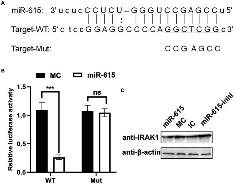
miR-615 targets IRAK1. (A) Bioinformatic prediction of the interactions between miR-615 and the 3′-UTR of swine IRAK1. For each schematic, the upper sequence is the sequence of mature miR-615, the middle sequence is the sequence in the binding site of miR-615 in the 3′-UTR of swine IRAK1, and the lower sequence is the mutated sequence of the IRAK1 3′-UTR. The seed sequence is underlined. (B) Luciferase activity in 293 T cells co-transfected with miR-615 mimics (or MC) and luciferase reporter gene plasmids containing the WT and Mut 3′-UTRs of IRAK1 for 48 h. Data are normalized against firefly luciferase activity. Comparisons between groups were determined using Student’s t-tests. ***p < 0.001, **p < 0.01. (C) The level of the IRAK1 protein during transfection with miR-615 mimics or inhibitors in IECs detected using western blotting during PEDV infection. Western blotting was conducted using anti-IRAK1 antibody at 24 h post-infection (hpi).

