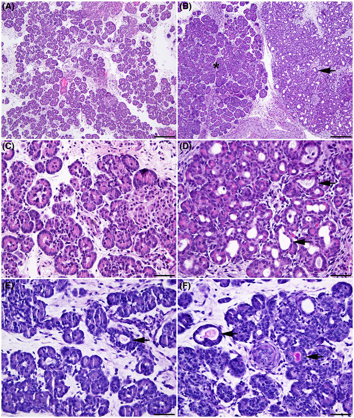
FIGURE 1.

Pancreatic pathology is evident by 80 days gestation in CFTR −/− sheep. Wild‐type (WT) (A, C, E) and CFTR −/− (B, D, F) sheep fetal pancreas at 80 days gestation. Normal pancreatic histology in the WT fetus (A). In the CFTR −/− fetus (B) both normal acini (asterisk) and an area with substantial acinar dilation (arrow) are adjacent to each other. Higher magnification images show normal pancreatic tissue in a WT fetus (C), and dilatation of multiple pancreatic acini (arrow) in a CFTR −/− fetus (D). Periodic Acid Schiff (PAS) staining of a normal pancreas in a WT fetus (E) and CFTR −/− pancreas (F). Note that occasional dilated acini are present in the WT pancreas, but no PAS‐staining material is visible in the lumen (arrow). In the CFTR −/− pancreas some dilated acini contain PAS‐staining material stains consistent with mucus. Hematoxylin and eosin stain (A to D). 100× (A and B); bar = 200 μm. 400× (C and D); bar = 50 μm. Period acid‐Schiff (PAS) stain (E and F). 400×; bar = 50 μm.
