FIGURE 2.

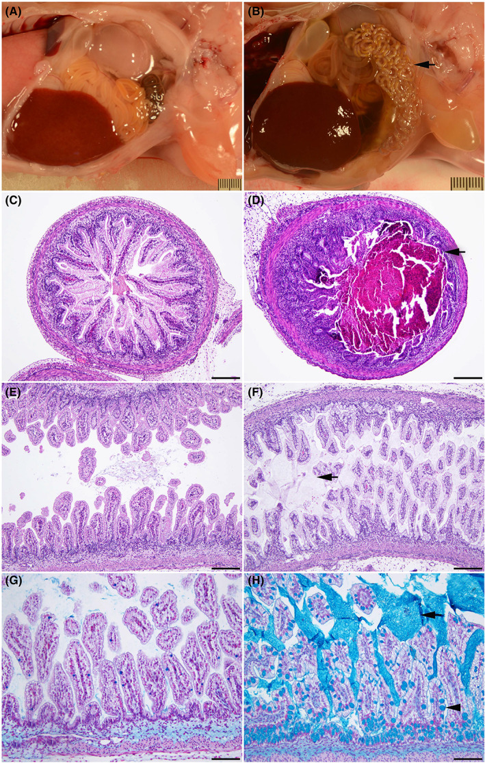
Intestinal obstruction is seen by 80 days gestation in the CFTR−/− sheep. WT (A, C, E, G) and CFTR −/− (B, D, F, H) sheep intestinal tracts at 80 days gestation. (A) Normal intestinal tract in a WT fetus. Note the constant diameter along the length of the intestine. (B) Early meconium ileus in a CFTR −/− fetus. Note the smaller diameter of a long segment of the small intestine and colon (arrow). (C) Normal small intestine in a wild‐type fetus. (D) Small intestine filled with meconium (arrow) in a CFTR −/− fetus. (E) Normal colon in a wild‐type fetus. (F) Colon with distended goblet cells in the mucosa and excessive luminal mucus (arrow) in a CFTR −/− sheep fetus. Note: hematoxylin and eosin (H and E) stain does not clearly stain the mucus. (G, H) Higher magnification images showing Alcian blue staining of mucus. A minimal amount of luminal mucus is seen in the WT colon (G). A large amount of luminal mucus (arrow) is evident in the CFTR −/− colon (H), where the goblet cells are also distended with mucus (arrowhead). H and E stain (A to F). 100× (C to F); bar = 20 μm. Alcian blue stain (G and H). 200× (G and H); bar = 100 μm.
