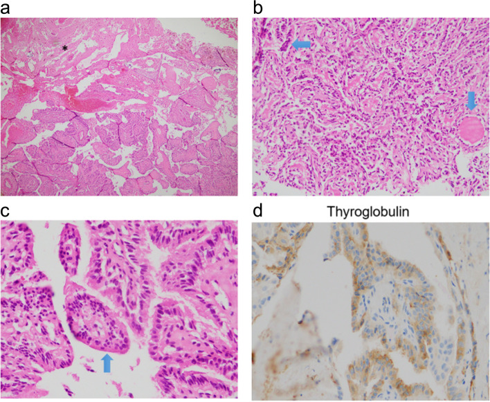
Fig. 8.
Histopathological and immunohistochemical images of brain metastases Histological images of metastatic papillary thyroid carcinoma (PTC). a PTC invades brain parenchyma (1/3 upper left *) (HE stain, × 40), b Image suggestive of papillary structureon papillary structure (long arrow), a follicle lined cuboidal cells with dark nucleic regularly contains colloid (short arrow) (HE × 100). c The papillary structures are covered by tumor cells without obvious papillary nuclear features (HE stain, × 400). d Tumor cells are positive Thyroglobulin (Immunohistochemistry stain, × 400). The histological image showed the majority of papillary structures in the peripheral area of tumor includes only vague papillary structures with edematous, collagenous and fibrous stroma like papillary cores, intercalating secretory follicular structures that contain colloid,. No tumor necrosis was seen and mitosis was rare. Immunohistochemistrical stain shows that tumor cells are positive for Thyroglobulin, TTF1 maker. Ki67 positivity was very low. Nuclear features of papillary thyroid carcinoma were not clear but there was no evidence of poorly differenciated or anaplastic thyroid carcinoma. The histopathological features are consistent with a high-grade papillary thyroid carcinoma

