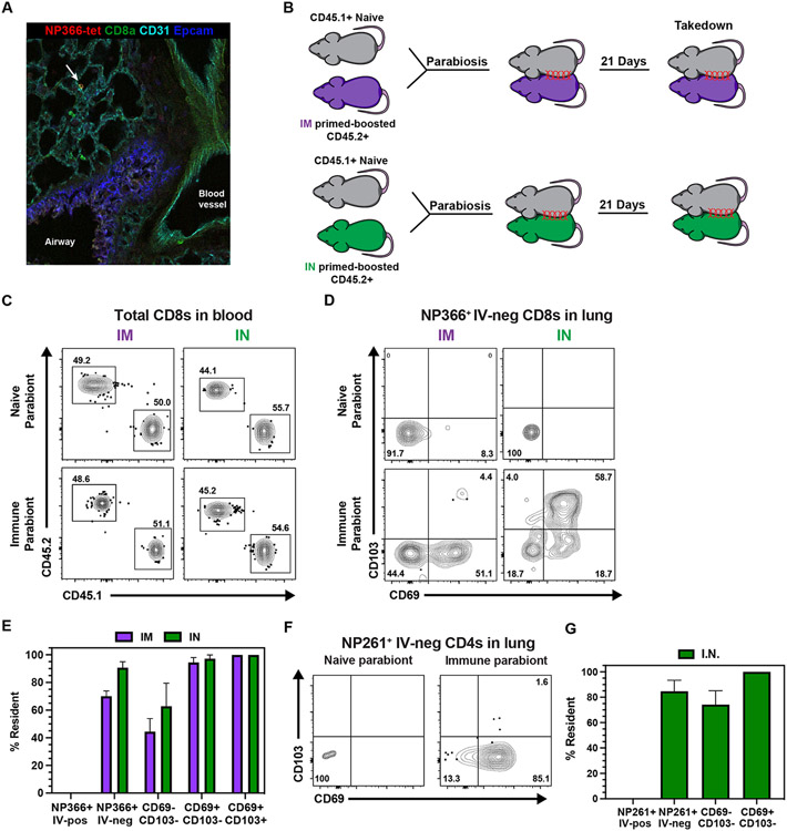
Fig 4. All mRNA immunization routes were sufficient to induce pulmonary resident memory CD8 T cells.
(A) Representative image of in situ NP366-tetramer staining in the lung upon IM prime-boost. (B) CD45.2+ mice were either IM or IN prime-boosted and then conjoined to CD45.1+ naïve hosts. After 21 days, lungs were harvested to examine recirculating and resident populations antigen-specific CD8 and CD4 T cells. (C) Representative flow cytometry plots depicting CD45.1+ and CD45.2+ CD8 T cells to demonstrate equilibration in the blood. (D-E) Representative flow cytometry plot (D) and quantification (E) of NP366-specific CD8 T cell subsets in the lung parenchyma. (F-G) Representative flow cytometry plot (F) and quantification (G) of NP261-specific CD4 T cell subsets in the lung parenchyma. Data represent N = 2 independent experiments with n = 4-5 mice per group, except for (A) which represents N = 1 independent experiment with n = 3 mice. Data are shown as mean ± SEM. *P<0.05, **P<0.01, ***P<0.001, ****P<0.0001as determined by unpaired two-tailed Student’s t test.

