Fig. 3.
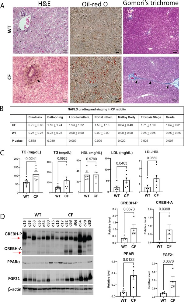
NASH phenotype and lipid disorder in CF rabbits. (A) Histological analysis of liver cellular structure (H&E, left column), lipid accumulation (oil-red O, central column), and collagen fiber (Gomori's trichrome staining, right column) in liver tissue sections from WT and CF rabbits. Arrows point to areas of hepatic inflammation or fibrosis. Scale bars: 20 μm in the left and middle panels; 100 μm in the right panel (B) Scoring for NASH activities in CF (n = 14, 50 ± 11 days of age) and WT (n = 4, 87 ± 3 days of age) rabbit livers based on the modified Brunt scoring system. (C) Levels of plasma total cholesterol(TC), triglycerides (TG), high-density lipoprotein (HDL), and lipoprotein cholesterol (LDL) in WT (n = 6, 50 ± 3 days of age) and CF (n = 6, 47 ± 3 days of age) rabbits. (D) Left: Western blot of Cyclic AMP-responsive element-binding protein H (CREBH), peroxisome proliferator-activated receptor α (PPARα) and Fibroblast growth factor 21 (FGF21) protein levels in WT and CF rabbits of different ages (indicated by numbers on the top of the gel, e.g., d15). Right: Quantification of CREBH, PPARα, and FGF21 protein levels in WT and CF rabbits at 50 to 70 days of age. CREBH precursor (CREBH-P): CREBH precursor; CREBH-A: activated CREBH protein.

