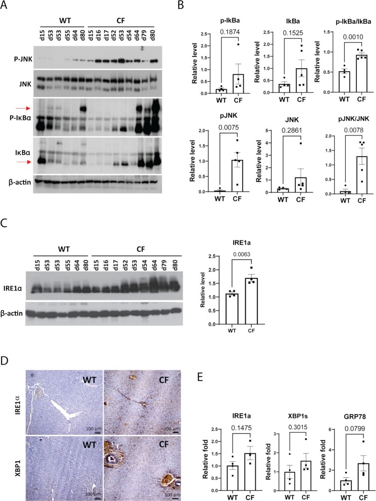
Fig. 5.
Inflammation and ER stress signaling in CF rabbit livers. (A) Western blot analyses of phosphorylated JNK (P-JNK), total JNK, phosphorylated IκB (P-IκB), and total IκB protein in WT and CF rabbits of different ages (indicated by numbers on the top of the gel, e.g., d15). (B) Quantification of Western blot results of phosphorylated JNK (P-JNK), total JNK, phosphorylated IκB (P-IκB) and total IκB protein in WT and CF rabbits at 50 to 70 days of age. (C) Left: Western blot analyses of IRE1α protein levels in WT and CF rabbits of different ages (indicated by numbers on the top of the gel, e.g., d15). Right: Quantification of Western blot results of IRE1α protein in WT and CF rabbits at 50 to 70 days of age. (D) IHC staining of IRE1α and XBP1 with liver sections from WT and CF rabbits of around 60 days old. Scale bars: 100 μm. (E) qPCR analyses of the mRNAs encoding ER stress sensor or mediators in the livers of WT and CF rabbits at 50 to 70 days of age.

