
Figure 3.
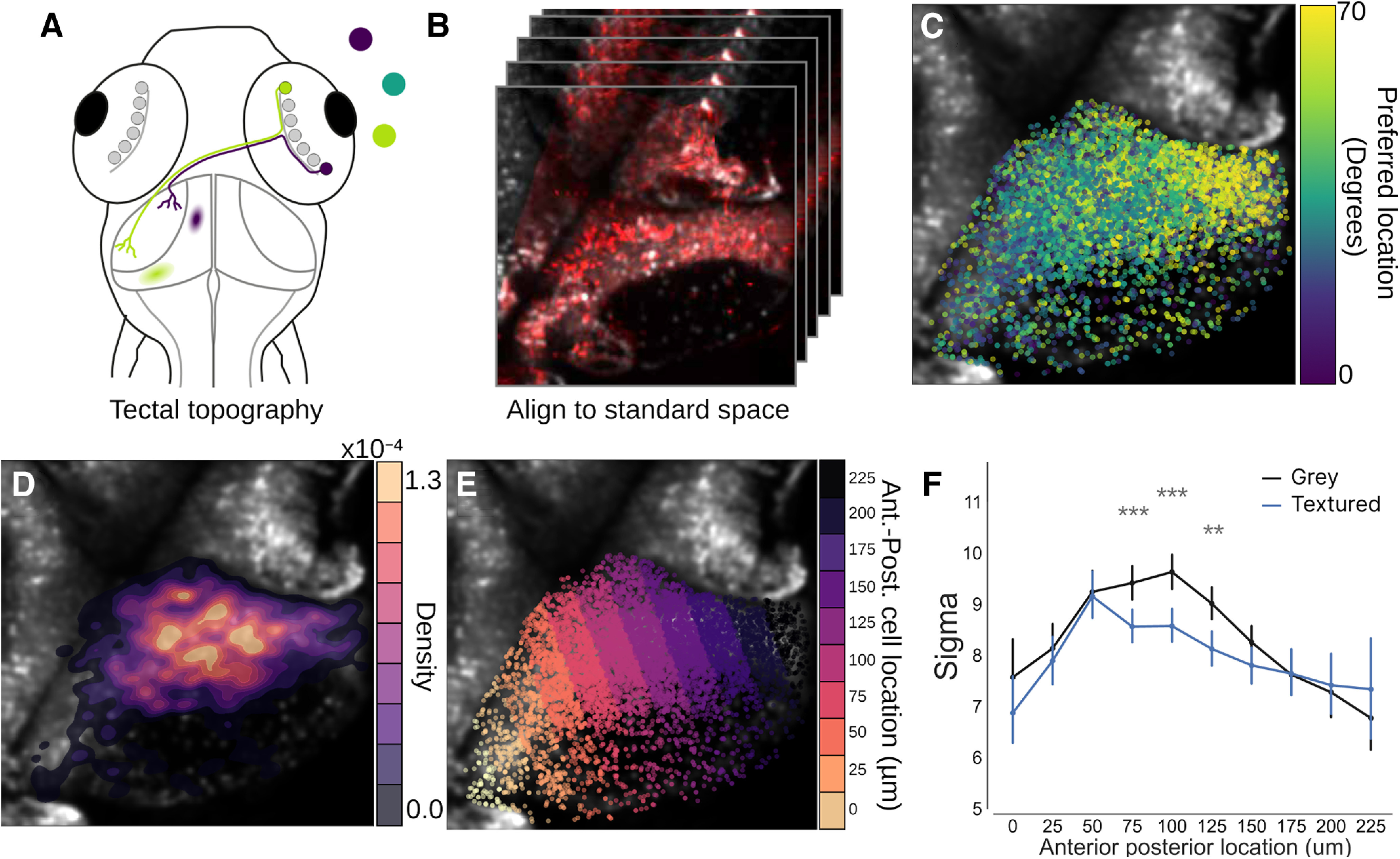
Modulated neurons are topographically distinct within the tectum. A, Schematic detailing the topographic organization of the tectum. Here, retinal ganglion cells project out of the retina and make synapses in the neuropil of the contralateral hemisphere. They do this is a way that preserves a spatial map of visual space within the tectum with frontal visual space mapping onto the anterior portion of the tectum (purple), whereas rear visual space maps more posteriorly (lime green). B, To assess the spatial arrangement of contextually modulated cells in the tectum a standard coordinate space was generated by aligning the functional imaging data to a high resolution stack of the tectum. C, Overlay of cells in the tectum which have been colored by their tuning preference to demonstrate the topography of the tectum. D, Overlay of a density heatmap showing the position of highly contextually modulated cells (Δ σ < −5) within the tectum. E, To quantify the position of contextually modulated cells, the tectum was divided into bins along its anterior-posterior axis. F, A plot of σ values for each segment within the anterior-posterior axis for both textured and gray backgrounds; **p < 0.01, ***p < 0.001.