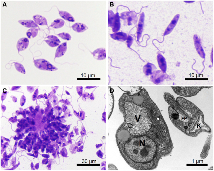
Figure 4.
Giemsa-stained Leishmania orientalis MHOM/TH/2021/CULE5 promastigotes from a culture of the patient’s biopsy tissue. (A) Procyclic form. (B) Leptomonad and nectomonad forms. (C) Rosette form. (D) Transmission electron micrograph showing the ultrastructure of the promastigotes. FP = flagellum within the flagellar pocket; K = kinetoplast; N = nucleus; V = large vacuole.

