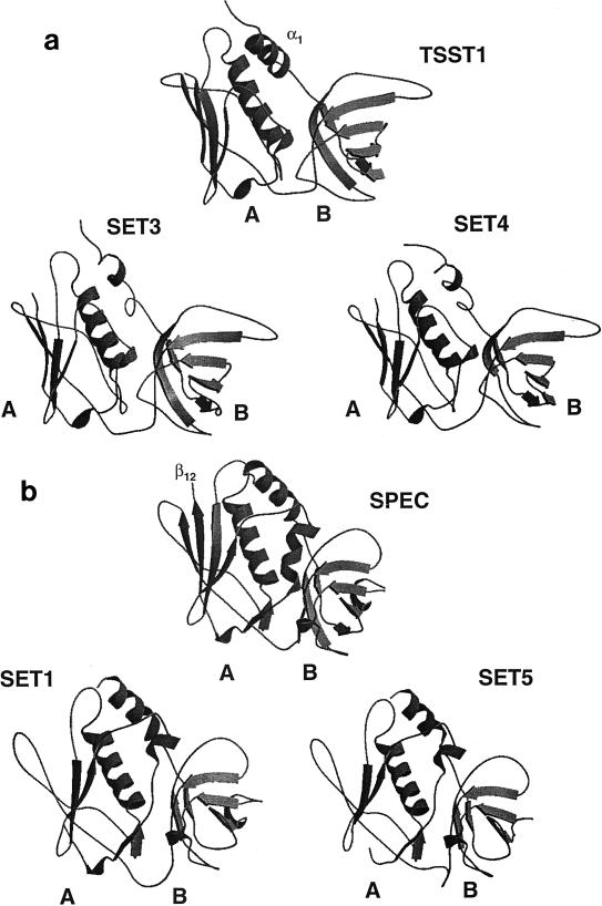
FIG. 3.
Schematic diagram of the modeled structures for SET proteins, TSST-1, and SPEC. The SET sequences were modeled against two target protein structures, TSST-1 (PDB file 2tss) and SPEC (PDB file 1an8) on a Silicon Graphics computer. All models showed the potential to form two domains: in domain A, the C terminus has a five-strand β-sheet surrounding a long central α-helix, and in the smaller domain B, the N terminus shows β-strands folding into a barrel creating the OB-fold. (a) The A domain of SET3 and SET4 has one less β-strand than the structure for TSST-1. (b) SET1 and SET5 differ from SPEC in having one less β-strand in the A domain. The missing strand corresponds to β12 in the SPEC model.

