Figure 5.

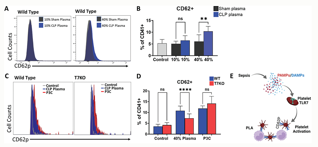
Septic plasma activates platelets and response is attenuated in platelets deficient of TLR7. Mice underwent sham (n = 2) or CLP surgery (n = 4) and 24 hours later whole blood was collected and processed to plasma. Platelets (n = 2) were isolated from whole blood of WT mice and treated with 10% or 40% (v/v) sham or CLP plasma. In the control group, same volume of Tyrode’s buffer was added. Activated platelets were analyzed using flow cytometry and defined as the percent of CD62P+ platelets over total platelets (CD62P+CD41+/CD41+x100%). (a) Representative flow cytometry histograms in WT platelets and (b) Corresponding bar graphs. Platelets isolated from whole blood of WT (n = 2) and T7KO (n = 2) mice were treated with 40% WT CLP plasma (n = 4) and CD62P+ platelets were quantified as above. In the control group, same volume of Tyrode’s buffer was added. (c) Representative flow cytometry histograms in WT and T7KO platelets treated with CLP plasma; (d) CLP plasma induced platelet activation in WT platelets and the response was attenuated in T7KO platelets. Samples treated with P3C (10 μg/ml) served as a positive control. (e) In sepsis, microbial infection leads to release of PAMPs and/or DAMPs that signals via platelet-TLR7 and contributes to downstream platelet activation and PLA formation. Created with BioRender.com. P3C = Pam3CSK4, T7KO = TLR7 deficient mice. WT = Wild-type. Bars represent the mean ± SD of 2 replicate experiments run with triplicate samples, **p< .01, ****p< .0001.
