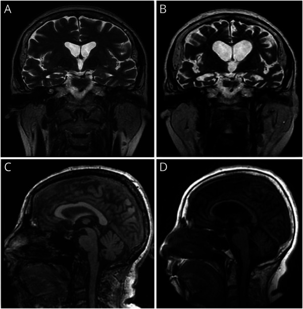
Figure 5. Brain Imaging Findings in a 63-Year-Old Woman With Progressive Functional Decline (Case 6).
Brain MRI coronal T2 images at ages 58 and 62 years (A and B) demonstrating progressive cortical atrophy in the frontotemporal region with progressive dilatation of the lateral and 3rd ventricles. Brain MRI midline sagittal FLAIR T1 images at ages 58 and 62 years (C and D) showing interval severe thinning and atrophy of the corpus callosum and atrophic changes in the midbrain and posterior fossa.

