. 2023 Jan 2;19(1):12–27. doi: 10.3988/jcn.2023.19.1.12

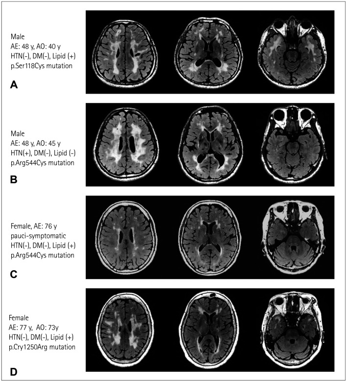
Fig. 1. Variable neuroimaging severity among patients with cerebral autosomal dominant arteriopathy with subcortical infarcts and leukoencephalopathy (CADASIL) carrying different NOTCH3 mutations. Representative fluid-attenuated inversion recovery (FLAIR) images showing (A) typical magnetic resonance imaging (MRI) features including lacunar infarct, diffuse white-matter hyperintensities (WMHs) involving the corona radiata, periventricular regions, and anterior temporal poles in a patient with CADASIL carrying a heterozygous NOTCH3 p.Ser118Cys mutation located within epidermal growth factor-like repeat (EGFR)2 of the NOTCH3 protein; (B and C) variable severity of WMHs without anterior temporal pole involvement in a middle-age-onset patient with CADASIL and an elderly paucisymptomatic individual, both of whom carried a heterozygous p.Arg544Cys NOTCH3 mutation that altered the amino acid residue between EGFR13 and EGFR14; and (D) typical MRI features of CADASIL in a late-onset patient with a heterozygous p.Cys1250Arg mutation located within the EGFR32. AE, age at examination; AO, age at stroke onset; HTN, hypertension; DM, diabetes mellitus; lipid, hypercholesterolemia.

Fig. 1