. 2023 Jan 2;19(1):12–27. doi: 10.3988/jcn.2023.19.1.12

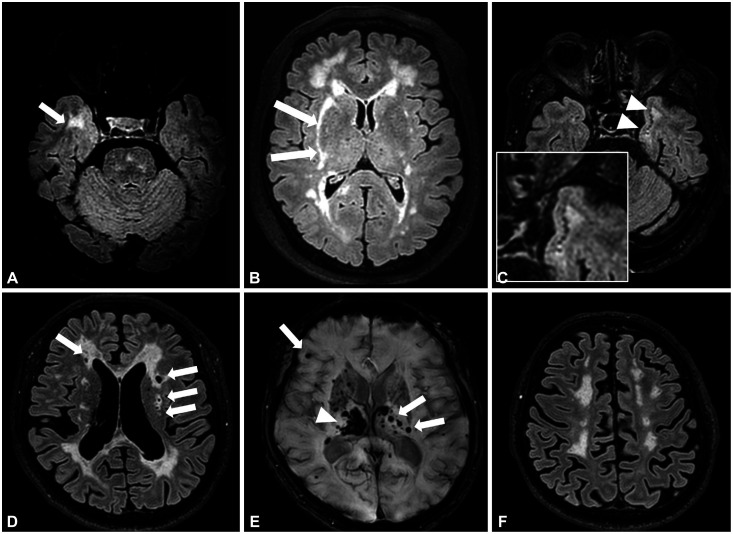
Fig. 3. Characteristic brain MRI features of CADASIL. Axial FLAIR MRI images of a 59-year-old male present prominent WMHs in the right anterior temporal lobe (arrow) (A). WMHs involving right external capsule (arrows) were noted in a 66-year-old female (B). Axial FLAIR MRI images of a 68-year-old male presented subcortical lacunar lesions at the junction between the white matter and the gray matter in the left anterior temporal lobe (arrowheads and the enlarged figure in the white box) (C). Multiple lacunar infarctions (arrows) at the bilateral subcortical white matter (D). Several cerebral microbleeds (arrows) with a right thalamic hematoma (arrowhead) (E) and marked brain atrophy (F). All patients had the same p.Arg544Cys mutation.

Fig. 3