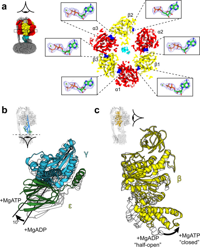
Fig. 2. Nucleotide occupancy and conformational changes in the F1 motor following incubation with MgATP.

a Horizontal section of the State 2 E. coli F1Fo ATP synthase cryo-EM map and details of the β subunit catalytic site occupancies (with equivalent mitochondrial F1 nomenclature:5 β1 = βDP, β2 = βE, β3 = βTP, as named for the E. coli enzyme16). β1 contains MgADP, β2 contains ADP, and β3 contains MgATP. Section of map contoured to 0.028 in ChimeraX62 and mesh for nucleotides contoured to isolevel 10 in PyMol (Schrödinger). b, c Comparison of the F1 motor after incubation with MgATP (this study; γ in blue, ε in green and β in yellow) or MgADP (PDB:6oqv19; subunits shown as outline). b The central stalk (subunits γ and ε) is rotated ~10° clockwise when viewed from the membrane (structures are aligned to the F1 β barrel crown). c β1 closes inwards from a half-open to closed conformation.
