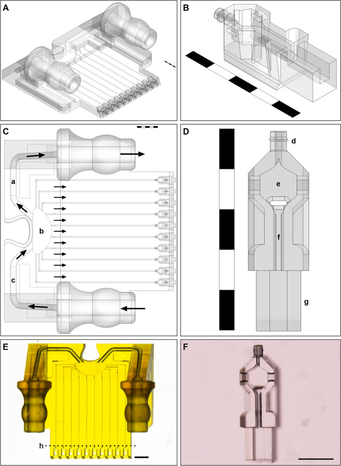
Figure 1.
Computer aided design (CAD) models of the nest and cradle (Scale bars A–E = 1 mm; F = 300 µm). (A) Isometric view of nest; (B) Isometric view of cradle; (C) Top view of nest (a—outlet channel, b—back reservoir, c—inlet channel, arrows indicate direction of fluid flow); (D) Top view of cradle (d—nozzle, e—cell chamber which is open at the top to enable cell manipulation, f—injection channel, g—fin); (E) Top image of nest at 1 × magnification on Nikon SMZ18 stereomicroscope. The dimensions of the nest ‘chip’ are 8.8 mm × 8.2 mm × 3.6 mm (nest printed at 10 × in adaptive resolution in course mode above line h and fine mode below line h); (F) Top image of cradle at 8 × magnification with a Nikon SMZ18 stereomicroscope (cradles printed at 20 × in course infill mode).

