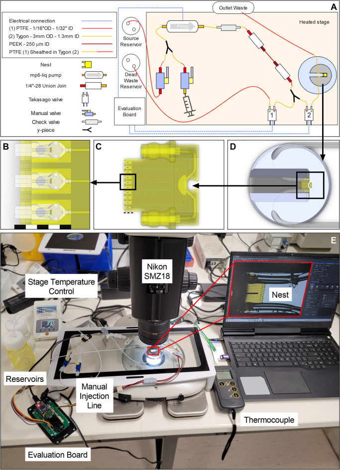
Figure 2.
Microfluidic testbed for nest-cradle device (Scale bars = 1 mm). (A) Diagrammatic representation of testbed components (components and positions are not to scale); (B) Zoom in on model of three central cradles inserted into the nest, which acts as the roof with cell loading holes for each cradle; (C) Zoom in on model of the whole nest with ten cradles inserted; (D) Model of nest with tubing attached secured by titanium holder in 60 mm petri dish; (E) Photographic image of testbed set up in the laboratory with a Nikon SMZ18 stereomicroscope. An Olympus IX83 inverted microscope was used in place of the SMZ18 for particle tracking velocimetry.

