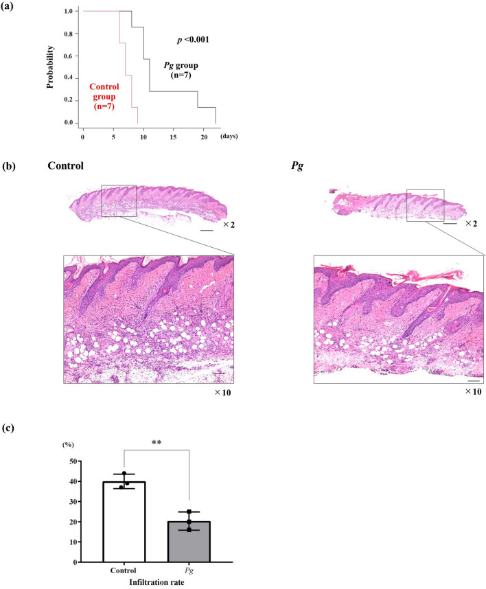
Figure 5.
Skin graft survival and pathological changes in skin grafts in both groups. (a) Skin graft survival between the groups (each group: n = 7). On day 8 after skin grafting, the degree of inflammation of the skin graft was evaluated pathologically (hematoxylin/eosin staining). (b) × 2 magnification. The bar scale is 500 µm, × 10 magnification The bar scale is 100 µm. (c) Percentage of inflammatory cell infiltration in both groups (each group: n = 3). *p < 0.05, **p < 0.005.

