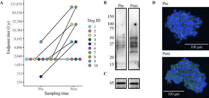
FIGURE 3.
Six of 10 dogs exhibited detectable antiglioma humoral responses. Humoral responses to vaccination were quantified using flow cytometry–based endpoint titers with disaggregated J3T spheroids (A). Representative Western blots (B/C) and confocal micrographs (D) depicting the serum antibody binding in a dog before (pre) immunization and 1-month post-vaccination (post). The Western blots show binding to proteins found in the vaccine lysate (serum from dog 7) with the lower blot (C) depicting β-actin loading control. The confocal images tested binding of antibodies to J3T spheroids (D; serum from dog 3). All serum samples used in the assays were obtained after two vaccinations (week 4).

