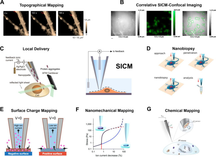
Figure 3.
SICM depicted in the center for constant-distance imaging using the ion current as a feedback signal. The principal biology applications and mapping modes are shown in panels A–G. (A) Topographical mapping of single cells, reproduced from Takahashi, Y.; Zhou, Y.; Miyamoto, T.; Higashi, H.; Nakamichi, N.; Takeda, Y.; Kato, Y.; Korchev, Y.; Fukuma, T. Anal. Chem.2020, 92 (2), 2159–2167 (ref (104)). Copyright 2020 American Chemical Society. (B) Correlative SICM-confocal imaging, reproduced with permission from Proceedings of the National Academy of Sciences USA Bednarska, J.; Pelchen-Matthews, A.; Novak, P.; Burden, J. J.; Summers, P. A.; Kuimova, M. K.; Korchev, Y.; Marsh, M.; Shevchuk, A. Proc. Natl. Acad. Sci. U.S.A.2020, 117 (35), 21637–21646 (ref (105)) under CC-BY 4.0 license. (C) Local delivery of biomolecules combined with laser-sheet microscopy. Reproduced from Li, B.; Ponjavic, A.; Chen, W. H.; Hopkins, L.; Hughes, C.; Ye, Y.; Bryant, C.; Klenerman, D. Anal. Chem.2021, 93 (8), 4092–4099 (ref (106)). Copyright 2021 American Chemical Society. (D) Single-cell nanobiopsy for cytoplasmic extraction. Reprinted from J. Biol. Chem.2018, Vol. 293, Toth, E. N.; Lohith, A.; Mondal, M.; Guo, J.; Fukamizu, A.; Pourmand, N. Single-cell nanobiopsy reveals compartmentalization of mRNAs within neuronal cells, 4940–4951 (ref (107)) under CC-BY 4.0 license. (E) Surface charge mapping in aqueous solution and (F) nanomechanical mapping. Reproduced from Clarke, R. W.; Novak, P.; Zhukov, A.; Tyler, E. J.; Cano-Jaimez, M.; Drews, A.; Richards, O.; Volynski, K.; Bishop, C.; Klenerman, D. Soft Matter2016, 12 (38), 7953–7958 (ref (108)) under CC-BY 3.0 license. (G) Extracellular pH mapping of single cells, reprinted by permission from Macmillan Publishers Ltd., Zhang, Y.; Takahashi, Y.; Hong, S. P.; Liu, F.; Bednarska, J.; Goff, P. S.; Novak, P.; Shevchuk, A.; Gopal, S.; Barozzi, I.; et al., Nat. Commun.2019, 10 (1), 5610 (ref (109)) under CC-BY 4.0 license.

