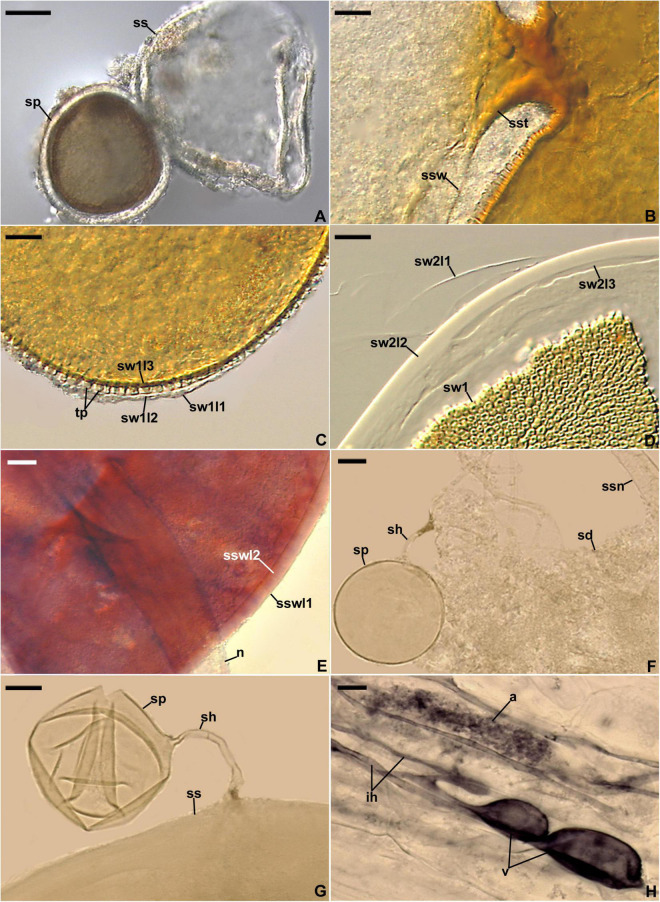
FIGURE 4.
Entrophospora infrequens. (A) Entrophosporoid morph with spore (sp) formed inside the sporiferous saccule (ss). (B) Funnel-shaped structure, continuous with spore wall 1 layer 3, supporting the sporiferous saccule wall (ssw). (C) Spore wall 1 layers (sw1l) 1–3; swl1 is almost completely sloughed off; tooth-shaped projections (tp) in cross-view are visible. (D) Spore wall 1 (sw1) and spore wall 2 layers (sw2l) 1–3. (E) Sporiferous saccule wall layers (sswl) 1 and 2, and the neck (n) of sporiferous saccule. (F) Juvenile glomoid spore (sp) with subtending hypha (sh) developed from the sporiferous saccule neck (ssn); soil debris (sd) are indicated. (G) Juvenile glomoid spore (sp) with subtending hypha (sh) developed from sporiferous saccule (ss). (H) Mycorrhiza with arbuscule (a), vesicles (v), and intraradical hyphae (ih). (A–D,F–H) Spores and mycorrhizal structures in PVLG. (E) Sporiferous saccule in PVLG+Melzer’s reagent. (A–H) Differential interference microscopy. Scale bars: (A) = 50 μm, (B–H) = 10 μm.

