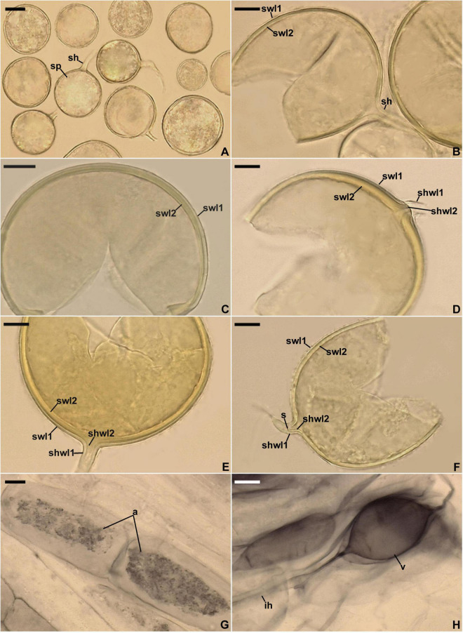
FIGURE 5.
Entrophospora infrequens. (A) Intact glomoid spores (sp) with subtending hyphae (sh). (B–F) Spore wall layers (swl) 1 and 2 continuous with subtending hyphal wall layers (shwl) 1 and 2; a septum (s) continuous with swl2 in the lumen of the sh is indicated in (F). (G,H) Arbuscules (a), intraradical hyphae (ih), and vesicles (v) in roots of Plantago lanceolata stained in 0.1% Trypan blue. (A–D,G,H) Spores and mycorrhizal structures in PVLG. (E,F) Spores in PVLG+Melzer’s reagent. (A–H) Differential interference microscopy. Scale bars: (A) = 20 μm, (B–H) = 10 μm.

