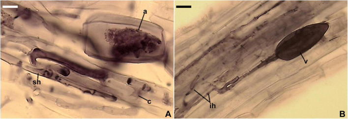
FIGURE 7.
Mycorrhizal structures of Entrophospora argentinensis in roots of Plantago lanceolata stained in 0.1% Trypan blue. (A) Arbuscule (a), coiled (c) and straight (sh) intraradical hyphae. (B) Vesicle (v) and intraradical hyphae (ih). (A,B) In PVLG. (A,B) Differential interference microscopy. Scale bars: (A) = 10 μm, (B) = 20 μm.

