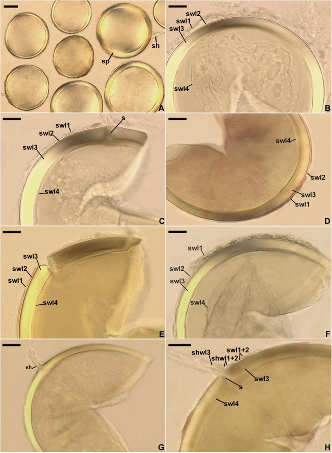
FIGURE 9.
Entrophospora furrazolae. (A) Intact spores (sp) with subtending hyphae (sh). (B–F) Spore wall layers (swl) 1–4; a septum (s) continuous with swl4 is indicated in (C). (G,H) Subtending hypha (sh) with subtending hyphal wall layers (shwl) 1–3; a septum (s) continuous with swl4 is indicated in (H). (A–C) Spores in PVLG. (D–H) Spores in PVLG+Melzer’s reagent. (A–H) Differential interference microscopy. Scale bars: (A) = 20 μm, (B–H) = 10 μm.

