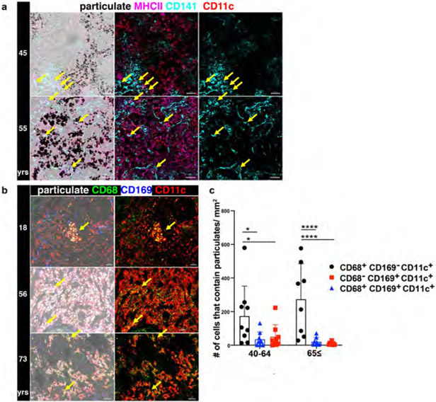
Extended Data Fig. 2. Particulate uptake occurs within CD11c+ macrophages but not dendritic cells (DC).
a. Confocal image of human LLNs stained for MHCII (HLA-DR), CD11c, and CD141 expression to identify DC (CD11c+HLA-DR+CD141+) shown with overlay of brightfield (left columns) for visualizing particulates (middle and right columns). Representative images were taken from 5 donors. Scale bar: 50 um. b. Confocal image of human LLNs stained for macrophage markers CD68 and CD169 along with CD11c. The image on the left show with overlay of brightfield (left columns) to show localization of particulates (white). The image on the right shows the CD68 and CD11c expression on macrophages. Representative images were taken from 6-9 donors per age group (≤39,40-64 and ≥65 yrs). Scale bar: 50-100 um. c. Graphs shows particulate content in specific macrophage subsets for different age groups, quantitated using Imaris software. Significance calculated by 2-way ANOVA with Tukey’s posttest, **P < 0.0021, ***P < 0.0002, ****P < 0.0001. Significance for 40-64: CD68+CD169−CD11c+ vs. CD68+CD169+CD11c+ P=0.0144, CD68+CD169−CD11c+ vs. CD68−CD169+CD11c+ P=0.0316; 65≤: CD68+CD169−CD11c+ vs. CD68+CD169+CD11c+ P=<0.0001, CD68+CD169−CD11c+ vs. CD68−CD169+CD11c+ P=<0.0001. Data presented as the mean ± SD. Data are from 17 donors.

