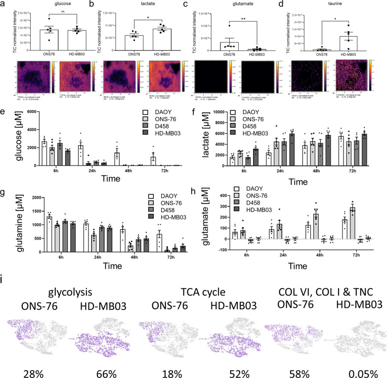
Fig. 2.
The 3D hydrogel models display subgroup-specific metabolic characteristics to those observed in MB patients. Several metabolites are known to be specifically high in MB patients [25]. 3D OrbiSIMS mass spectrometry imaging also confirms relatively high metabolite levels of glucose a. Lactate levels b are elevated in Group 3 nodules (D458, HD-MB03), while glutamate c is specifically high in SHH (DAOY, ONS-76). The Group 3-specific metabolite taurine d is also significantly higher in Group 3 nodules compared to SHH (a–d: mean ± SEM; n = 5; a, b: unpaired t-test, c: Mann Whitney test, d: unpaired t-test with Welch correction; *P < 0.05, **P < 0.01). Gel supernatants of 3 week-old SHH and Group 3 nodules were collected 6, 24, 48 and 72 h after medium change (containing 5000 µM glucose and 2000 µM glutamine) to measure consumption and secretion of glucose e, lactate f, glutamine g and glutamate h. Note that although both subgroups quickly consume glucose and glutamine, only lactate is secreted by both. Interestingly, glutamate secretion is characteristic for SHH nodules and below detection limit in Group 3 nodules (mean ± SEM; n = 3). i Gene expression for glycolysis (ALDOA, LDHB, GALM, AKR1A1, HKDC1, ENO2, ALDH2, ENO3, PGAM2, PFKM, PDHA1, PCK2, ALDH9A1), TCA cycle (SUCLG1, PDHA1, PCK2, MDH2, PC, MDH1, FH) and ECM genes (COL6A1, COL6A2, COL6A3, COL1A1, TNC) at the single cell level are displayed for the SHH (ONS 76) and Group 3 (HD MB03) model (purple: gene expressing cell; grey: non-expressing cell; percentage indicates relative proportion of total cells expressing the specified gene set). Cluster specific expression of these gene sets is shown in Additional file 2: Fig. S6

