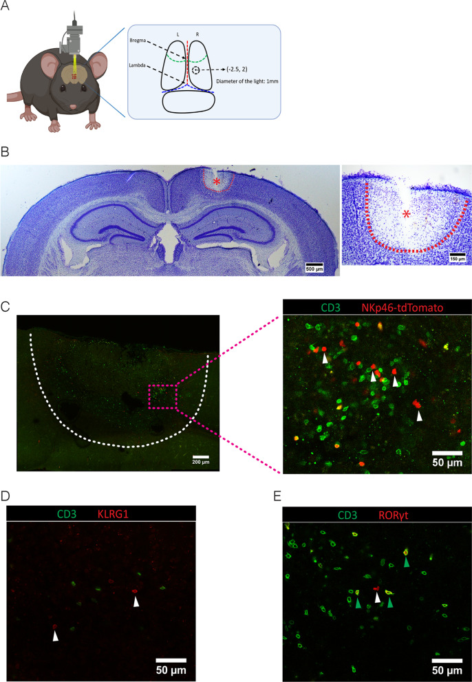
Fig. 1.
Presence of ILCs in the ischemic stroke lesion. A Coordinates of the lesion on the brain of photothromobotic (PT) mouse model at 2.5 mm posterior to bregma and 2.0 mm lateral to midline with 1 mm diameter. B Nissl staining on the vibratome section to indicate the lesion in the stroke brain at P2. Data represent n = 4 mice. C Immunofluorescence on the vibratome sections of stroke brain at P10. Since the markers of ILCs, NKp46, KLRG1 and RORγt are also expressed by CD3+ non-ILCs, we used CD3 to distinguish NK/ILC1s from NKT cells, ILC2s from regulatory T cells (Tregs) and NKp46+ ILC3s from Th17 cells. CD3−NKp46+ cells (NK, ILC1 or NKP46+ ILC3, white arrows). Data represent n = 4 mice. D CD3−KLRG1+ cells (ILC2, white arrows). Data represent n = 4 mice (E) CD3+RORγt+ cells (Th17, green arrows) and CD3−RORγt+ cells (ILC3, white arrows). Data represent n = 4 mice

