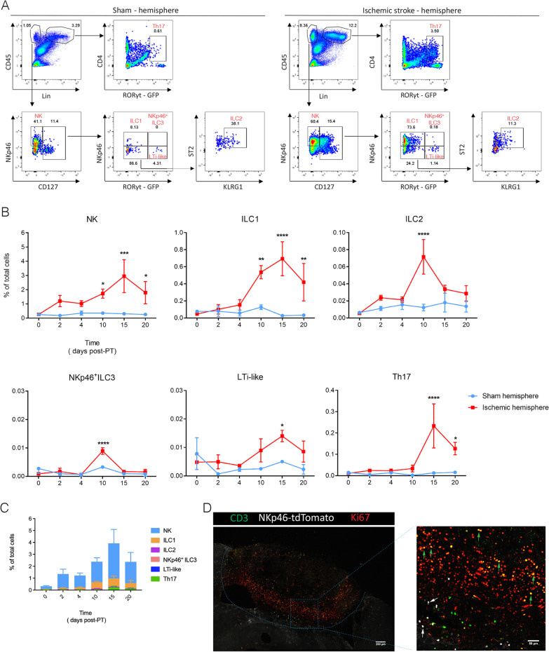
Fig. 2.
Time-course of ILC migration towards the ischemic stroke lesion. Cytometry analysis of ILCs in the sham (A, left) and P15 stroke hemisphere (A, right), NK (CD45+Lin−NKp46+CD127−), ILC1 (CD45+Lin−NKp46+CD127+RORγt−), ILC2 (CD45+Lin−NKp46−CD127+RORγt−ST2+KLRG1+), NKp46+ ILC3 (CD45+Lin−NKp46+CD127+RORγt+), LTi-like (CD45+Lin−NKp46−CD127+RORγt+), and Th17 (CD45+Lin+CD4+RORγt+). Representative dot plots of ILCs and Th17 cells on P15 are shown with the averages for the gates. Each panel is representative of 3 independent experiments (n = 3 mice). (B) Quantification of ILC populations and Th17 cells in the hemisphere after PT induction. The percentage of each population in total live cells (named total cells) was calculated. Data represent n = 3 mice. NK: P10 *P = 0.0364, P15 ***P = 0.0003, P20 *P = 0.021; ILC1: P10 **P = 0.0052, P15 ****P < 0.0001, P20 **P = 0.008; ILC2: P10 ****P < 0.0001; NKp46+ ILC3: ***P = 0.0001; LTi-like: P15 *P = 0.0232; Th17: P10 ****P < 0.0001. C Comparison of ILC and Th17 cell numbers in stroke hemisphere at different time points after induction. Data represent n = 3 mice. D Confocal images showing immunofluorescence for CD3, NKp46 and Ki67 in the PT-induced lesion at day 10. Green arrows point to the proliferating CD3+ cells, while white arrows point to the non-proliferating CD3−NKp46+ cells including NK cells, ILC1s and ILC3s. Data represent n = 2 mice

