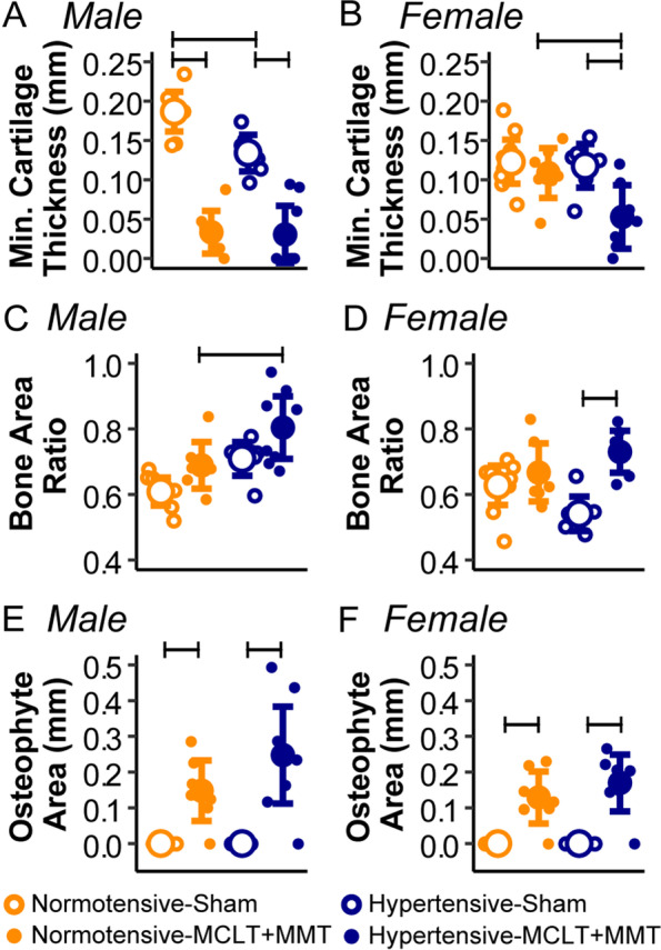
Fig. 3.

Quantitative histological results for male (left) and female (right) tibial region in the medial joint compartment. A In males, minimum cartilage thickness decreased with MCLT+MMT compared to sham in both hypertensive and normotensive animals. B In females, decreased cartilage thickness due to MCLT+MMT only occurred in hypertensive animals. C The ratio of the bone area associated with the subchondral bone plate to the total area of bone and trabecular bone in the subchondral space (“bone area ratio”) increased in hypertensive-MCLT+MMT males compared to normotensive-MCLT+MMT males. D Hypertensive-MCLT+MMT females also had an increased bone area ratio; however, this was compared to hypertensive-sham animals. E, F Osteophyte areas increased due to MCLT+MMT in both strains and sexes. Bars represent p < 0.05 between the groups. Data are presented as mean ± 95% confidence interval
