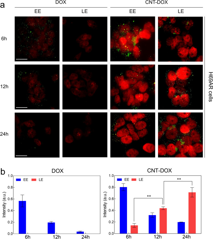
Fig. 4.
Intracellular trafficking of nanodrugs. a Confocal microscopy images visualizing DOX intensity (red) in the nuclei of H69AR cells and the indicated vesicles (early endosomes (EEs) and late endosomes (LEs), green) after treatment with free DOX and the CNT-DOX for 6, 12 and 24 h. The scale bar is 75 μm. b Time-dependent fluorescence intensity of EE and LE after treatment with free DOX and CNT-DOX for the indicated times (6, 12, and 24 h). The fluorescence intensities of LE were only found after incubating with CNT-DOX for 6, 12, and 24 h and analyzed by normalizing the fluorescence intensities of confocal images. All data represent the mean ± SEM (n = 6). *p < 0.05, **p < 0.01 and ***p < 0.001

